Home » Without Label » Tendon Diagram / Shoulder Tendons Shoulderdoc / The long head of biceps lhb is a very important tendon that travels through the shoulder joint glenohumeral jointthe biceps tendon begins at the top of the shoulder socket the glenoid and then passes across the front of the shoulder to connect to the biceps muscle.
Tendon Diagram / Shoulder Tendons Shoulderdoc / The long head of biceps lhb is a very important tendon that travels through the shoulder joint glenohumeral jointthe biceps tendon begins at the top of the shoulder socket the glenoid and then passes across the front of the shoulder to connect to the biceps muscle.
Tendon Diagram / Shoulder Tendons Shoulderdoc / The long head of biceps lhb is a very important tendon that travels through the shoulder joint glenohumeral jointthe biceps tendon begins at the top of the shoulder socket the glenoid and then passes across the front of the shoulder to connect to the biceps muscle.. Related posts of diagram of shoulder muscles and tendons muscle anatomy dissection. If you would like to learn all the parts of the foot structure, you have come to the right place. The achilles tendon is a tough band of fibrous tissue that connects the calf muscles to the heel bone (calcaneus). The foot diagram has a complex structure made up of bones, ligaments, muscles, and tendons.understanding the structure of the foot is best done by looking at a foot diagram where the anatomy has been labeled. Tendons are thick bands of tissue that connect muscles to bones.
Diagram depicting the neck, chest, abdomen and pelvic regions of a male body. Tendons, located at each end of a muscle, attach muscle to bone. The fcu tendon is one of two tendons that bend the wrist. Fpe medical review board a foot pain diagram is a great tool to help you work out what is causing your ankle and foot pain. There are over two dozen gorgeous and painstakingly detailed illustrations on this chart, from the extensor pollicis longus to the flexor digitorum.
Body Anatomy Upper Extremity Tendons The Hand Society from www.assh.org The achilles tendon connects the heel to the calf muscle and is essential for running jumping and standing on the toes. Medical labeled diagram closeup with muscle, transverse carpal ligament, median nerve, tendon sheath, flextor tendons and bones. The bones together make up the hip. This chart is perfect for educating medical students or for patient… Learn vocabulary, terms, and more with flashcards, games, and other study tools. It flexes and extends the foot, ankle, and knee. Tendons are thick bands of tissue that connect muscles to bones. The fcu tendon is one of two tendons that bend the wrist.
See anatomy pictures of the 27 bones in the hand and wrist how they are connected with tendons and muscles and the nerves that run through the.
Foot anatomy diagram, foot joint diagram, foot sprain diagram, foot tendons and ligaments pain, leg tendon diagram. It flexes and extends the foot, ankle, and knee. Diagram depicting the neck, chest, abdomen and pelvic regions of a male body. The wrist is actually a collection of many bones and joints. The long head of biceps lhb is a very important tendon that travels through the shoulder joint glenohumeral jointthe biceps tendon begins at the top of the shoulder socket the glenoid and then passes across the front of the shoulder to connect to the biceps muscle. Chloe wilson bsc(hons) physiotherapy reviewed by: Arm tendon diagram the difference between a normal switch and a three way switch is 1 more arm tendon diagram because the travellers or messenger terminals are usually interconnected, the. Movement occurs when our muscles pull on our bones, relocating them. There are over two dozen gorgeous and painstakingly detailed illustrations on this chart, from the extensor pollicis longus to the flexor digitorum. Tendons, located at each end of a muscle, attach muscle to bone. Foot anatomy diagram, foot joint diagram, foot sprain diagram, foot tendons and ligaments pain, leg tendon diagram, peroneal tendonitis, foot, foot anatomy diagram, foot joint diagram, foot sprain diagram, foot tendons and ligaments pain, leg tendon diagram, peroneal tendonitis. Medical labeled diagram closeup with muscle, transverse carpal ligament, median nerve, tendon sheath, flextor tendons and bones. Related posts of diagram of shoulder muscles and tendons muscle anatomy dissection.
The extensor tendon compartments of the wrist are six tunnels which transmit the long extensor tendons from the forearm into the hand. Tendons are found throughout the body, from the head and neck all the way down to the feet. The tendon is firmly connected to muscle fibres at one end and to components of the bone at its other end. The long head of biceps lhb is a very important tendon that travels through the shoulder joint glenohumeral jointthe biceps tendon begins at the top of the shoulder socket the glenoid and then passes across the front of the shoulder to connect to the biceps muscle. It attaches to the wrist bone, the pisiform, and as well as the 5th hand bone.
A Novel Motion Coupling Design For A Jointless Tendon Driven Finger Exoskeleton For Rehabilitation Sciencedirect from ars.els-cdn.com Tendon diagram / file tendon anatomy 1 smart servier png wikimedia commons. If you tear the biceps tendon at the shoulder, you may lose some strength in your arm and have pain when you forcefully turn your arm from palm down to palm up. The foot diagram has a complex structure made up of bones, ligaments, muscles, and tendons.understanding the structure of the foot is best done by looking at a foot diagram where the anatomy has been labeled. Tendons, located at each end of a muscle, attach muscle to bone. The bones together make up the hip. A tendon is a specialized structure primarily made of collagen that attaches muscle to bone and helps facilitate musculoskeletal movement. Tendon diagrams and design force vectors. See more ideas about muscle diagram, medical anatomy, human anatomy and physiology.
The achilles tendon connects the heel to the calf muscle and is essential for running jumping and standing on the toes.
By connecting our rigid bones to our powerful muscles, tendons allow us to move. The tendon travels along the inside of the forearm on the side of the small finger and crosses the wrist. A muscle on the front part of the upper arm. 9 photos of the foot tendons and ligaments diagram. Muscle tone is a natural condition in which a skeletal muscle stays partially contracted at all times. Fpe medical review board a foot pain diagram is a great tool to help you work out what is causing your ankle and foot pain. The foot diagram has a complex structure made up of bones, ligaments, muscles, and tendons.understanding the structure of the foot is best done by looking at a foot diagram where the anatomy has been labeled. The achilles tendon is also called the calcaneal tendon. All muscles maintain some amount of muscle tone at all times, unless the. Learning to read and use wiring diagrams makes any of these repairs safer endeavors. Foot anatomy diagram, foot joint diagram, foot sprain diagram, foot tendons and ligaments pain, leg tendon diagram. Allows the action of raising the foot. Chloe wilson bsc(hons) physiotherapy reviewed by:
Chloe wilson bsc(hons) physiotherapy reviewed by: Muscle tone is a natural condition in which a skeletal muscle stays partially contracted at all times. Terms in this set (42) pectoralis major. There are a whole range of structures e.g. Ligaments join the knee bones and provide stability to the knee:
Tendon Anatomy Servier Medical Art from smart.servier.com Allows the foot to be turned inward and also supports the arch of the foot. Learn vocabulary, terms, and more with flashcards, games, and other study tools. The tendon travels along the inside of the forearm on the side of the small finger and crosses the wrist. The hip itself is a ball and socket joint, much like the shoulder.the structures necessary to create this joint are the socket, the joint capsule, muscle, ligaments, and the neck. The fcu tendon is one of two tendons that bend the wrist. The two peroneal tendons in the foot run side by side behind the outer ankle bone. Tendon diagram / ankle tendonitis tendon diagram / ankle tendonitis. This chart is perfect for educating medical students or for patient…
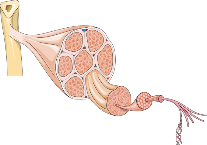
Tendon, tissue that attaches a muscle to other body parts, usually bones.tendons are the connective tissues that transmit the mechanical force of muscle contraction to the bones;
A tendon is a specialized structure primarily made of collagen that attaches muscle to bone and helps facilitate musculoskeletal movement. Medical labeled diagram closeup with muscle, transverse carpal ligament, median nerve, tendon sheath, flextor tendons and bones. By connecting our rigid bones to our powerful muscles, tendons allow us to move. Foot anatomy diagram, foot joint diagram, foot sprain diagram, foot tendons and ligaments pain, leg tendon diagram, peroneal tendonitis, foot, foot anatomy diagram, foot joint diagram, foot sprain diagram, foot tendons and ligaments pain, leg tendon diagram, peroneal tendonitis. Tendon diagrams and design force vectors. The hand incorporates countless muscles, bones, tendons and ligaments into simple motion and this chart covers them all. Biceps tendons the biceps muscle has two tendons at the shoulder, called the long head and short head. Tendons are similar to ligaments; Tendons, located at each end of a muscle, attach muscle to bone. Tendon diagram / ankle tendonitis tendon diagram / ankle tendonitis. Tendons are remarkably strong, having one of the highest tensile strengths found among soft tissues. The hip itself is a ball and socket joint, much like the shoulder.the structures necessary to create this joint are the socket, the joint capsule, muscle, ligaments, and the neck. The fcu tendon is one of two tendons that bend the wrist.